MR Arthrography

MR Arthrography
Since MRI has been considered as the best tool to investigate the joints, the radiologists have always been concerned with the possibility of obtaining more elaborate information to aid the surgeon during the operation and to predict the incidents before it. As far as the whole components of the joints would not be discerned by MRI, MR Arthrography would be applied to provide clearer images of the malfunctioning joints. MR Arthrography works by expansion of the joint space with a diluted contrast agent.
Thus the intended image is obtained by separating the adjacent components. This method is the optimum one with the lease harmful consequences. Moreover, it is minimally invasive and requires sterile conditions along with certain precautions.
MR Arthrography could be applied on shoulder joints to investigate glenohumeral, humral legaments and labrum ligaments. It also could be applied to investigate and to separate the post-surgery lacerations of the meniscus, or to check the labrum injuries of the pelvises.
It depends on the radiologist’s skill to perform it well. It could be done under the guide of fluoroscopy, sonography, CT scan or MRI.
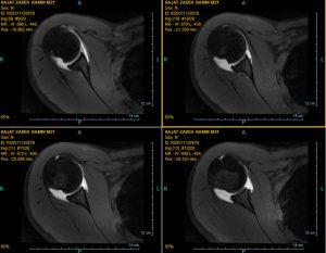
A 21-year-old young man visited the imaging center quite a while ago. He was seeking the exemption of the compulsory military service. In MRA that I did ALPSA and Hill socks lesions are seen. Though, the latter could be seen through a general MRI.

