4. 4D Sonography
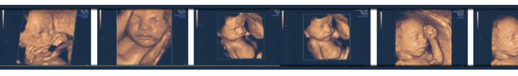
3D and 4D are most of the time being applied in embryonic and fetal studies. 2D is the commonest in use in which the movements of the fetus are represented on the screen. These waves are ultrasound waves. This type of imaging is being applied to analyze the health of the embryos. If the waves are sent through different directions, the recreated computerized images are 3D and 4D. the difference between these two is the capability to see the embryo’s movement in 4D. the movements are definitely shown with some time lapses. These occur due to the time required for the computer to do the computing process. Therefore, 4D is more expensive than 3D. it requires high speed computers and some specific probes.
Hence, there had been no specific side effects for 3D or 4D imaging, it is truly recommended that they should be prescribed by an expert only. The types of sonographies either 2D, 3D or 4D are similar in nature and the differences are the time, the strength of the waves and the intervals. Therefore:
- It should be less than 30 minutes, however, the safest time limit hasn’t been designated for it.
- The strength of the waves should not exceed FDA standards, however, the modern sets are already adjusted no to exceed the limits.
- It is recommended that it should not be done more than once a month(i.e. as least as possible).
As long as the mothers get stressed out during this type of imaging which reveals clear and perceptible images, it is recommended that 3D and 4D should be done later than the first 17 weeks of pregnancy. Although they provide confidence for the mothers as their main task, they should not be the source of harm whatsoever.
26th to 30th weeks of pregnancy are so ideal for such imaging. Though before or after such period has been advised. It should be mentioned that from 32nd week of pregnancy this type of sonography becomes almost impossible due the amniotic fluid reduction.
It is recommended to drink sufficient beverages a week prior if possible to increase the fluid level.
Further usage:
.one of the other reasons concerning such sonographies are to check the process of ovulating to occur at a definite proper area within the uterus at the IVF.
.to locate the intervention in order to check the peripheral nerves. Recently such method has been applied to check the anal sphincter laceration, its position, size and extension. And also perineum for the first timers.
Ultrasound focused shock wave therapy: is done for the first time in Iran. It is a non-invasive method to treat without applying medicine. It could be applied for problems such as heels spurs, tennis elbow, Achilles tenopathy, planter fasciitis, calcified tendinitis of shoulder, hypertrophic and atrophic non-union of bones, as well foot ulcers for those suffering diabetes.

